

Removal of Benign Skin Lesions
Moles, Keratoses, and Benign Skin Lesions
Medical Removal Options
Targeted medical approaches to precisely remove benign skin lesions while minimizing scarring and promoting fast healing.
CMQ-CERTIFIED PHYSICIANS · NATURAL RESULTS · SERVICE IN FRENCH AND ENGLISH
to know
What are Benign Skin Lesions?
Over time, the skin can develop various benign skin lesions on the face, neck, or body. Although generally harmless, these lesions can be cosmetically bothersome, get irritated by clothing, cause discomfort, or raise concerns about their evolution.
The most frequently encountered lesions include cherry angiomas (red vascular spots), keratoses (skin thickenings related to age or sun exposure), skin tags (acrochordons), and moles (navi). Each type of lesion has specific characteristics and requires a tailored removal approach.
A prior medical evaluation is essential and mandatory before any removal — to confirm the benign nature of the lesion, ensure the absence of suspicious signs, and determine the most appropriate and safest removal method based on the type, location, and depth of the lesion.
Addressed Concerns
- Cherry Angiomas (red spots)
- Seborrheic Keratoses
- Actinic Keratoses
- Skin Tags (acrochordons)
- Moles (benign nevi)
- Other confirmed benign lesions
Treatment options
Types of Lesions & Treatment options
Four common types of benign lesions, each with its specific medical removal approach.
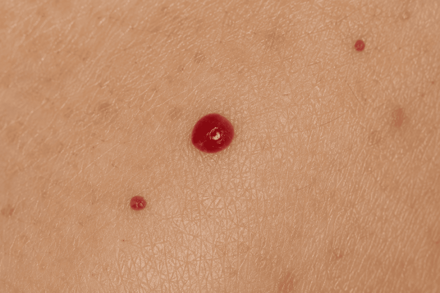
Cherry Angiomas (Red Spots)
Benign Vascular Lesions
Cherry angiomas are small red growths formed by clusters of dilated blood vessels. Laser treatment targets hemoglobin in the vessels to reduce the lesion with minimal impact on surrounding skin.
- Small red spots on the face or body
- Precise laser targeting of blood vessels
- Minimal impact on surrounding skin
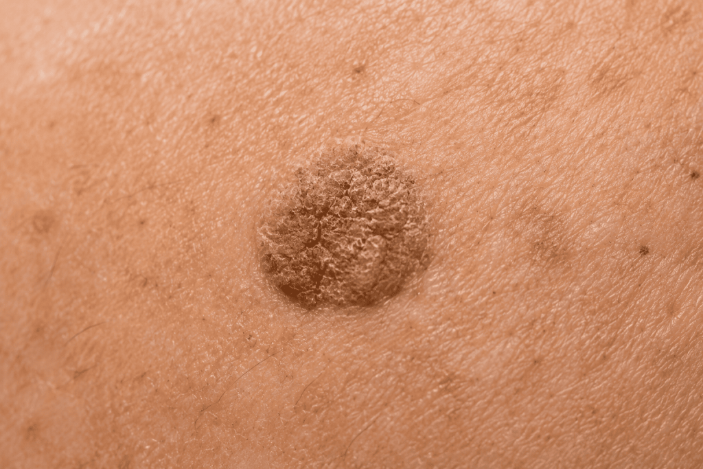
Keratosis
Keratinic Growths
Keratoses — seborrheic or actinic — are thickenings of the outermost layer of the skin. The laser allows precise ablation, promoting more even skin after healing.
- Seborrheic Keratoses
- Precise laser ablation of the lesion
- More even skin after healing
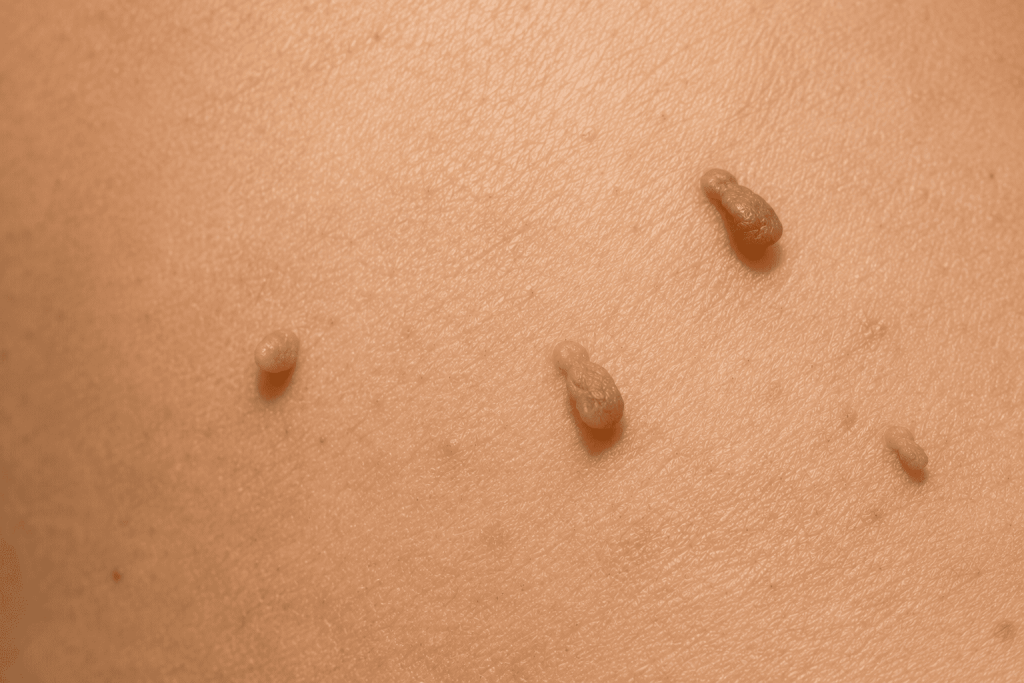
Skin Tags (Acrochordons)
Small Pedunculated Skin Growths
Skin tags are small skin-colored growths attached to the skin by a thin stalk, common on the neck, eyelids, and folds. Laser removal is targeted and generally well-tolerated.
- Neck, eyelids, armpits, and skin folds
- Targeted and precise laser excision
- Generally fast healing
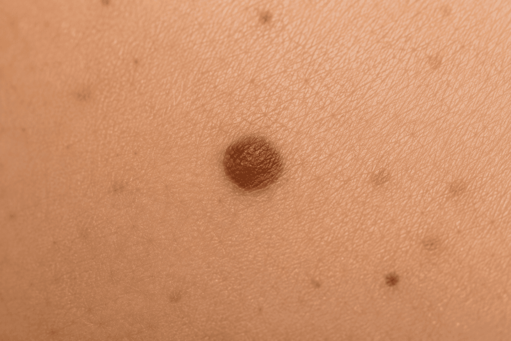
Moles (benign nevi)
Benign Pigmented Lesions
Moles are clusters of pigmented cells that can be flat or raised. A prior medical evaluation is mandatory to confirm their benign nature before any removal.
- Mandatory medical evaluation prior to removal
- Laser removal or another adapted technique
- When medically confirmed
Our Recommendation
Choosing the most appropriate approach
The choice of removal method depends on several factors that our doctors will evaluate during your mandatory medical consultation:
Nature and type of lesion
Location on the face, neck, or body
Size and depth
Your phototype and skin sensitivity
Your aesthetic goals and desired recovery time
A medical consultation is mandatory before any skin lesion removal — to confirm the benign nature of the lesion and determine the safest and most appropriate approach for your situation.
Results
Before & After
Real patient results from Medispa Physimed.
These photos are provided for informational purposes only and do not constitute a guarantee of results
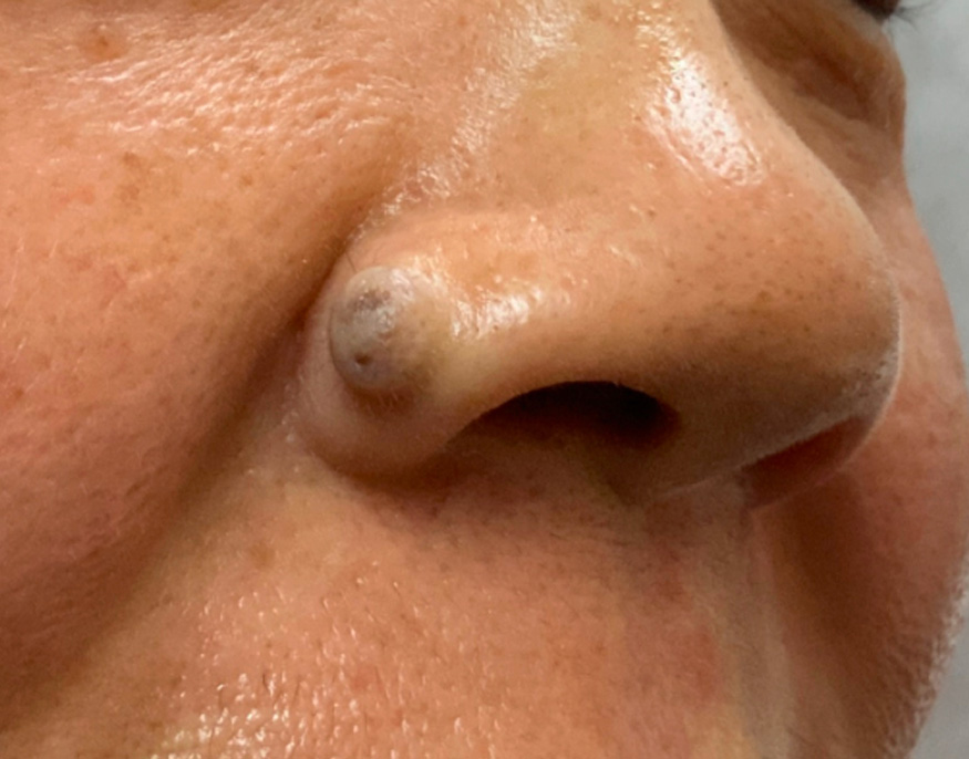 BEFORE
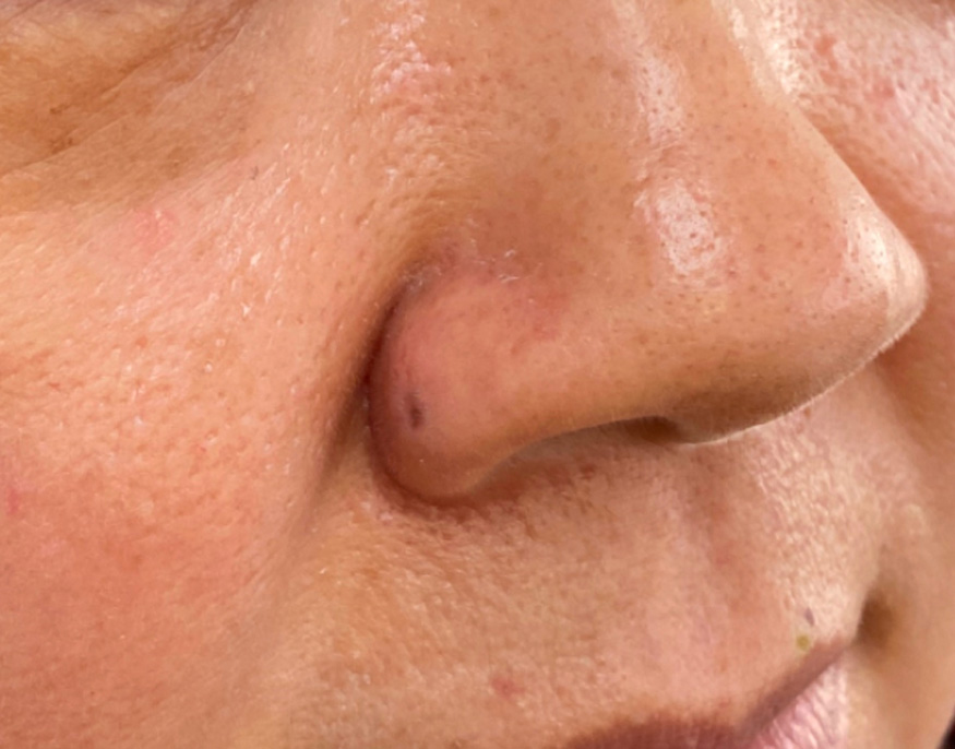 AFTER
AFTER
Keratosis
Remove benign skin lesions with precision, minimizing scarring and promoting rapid healing.
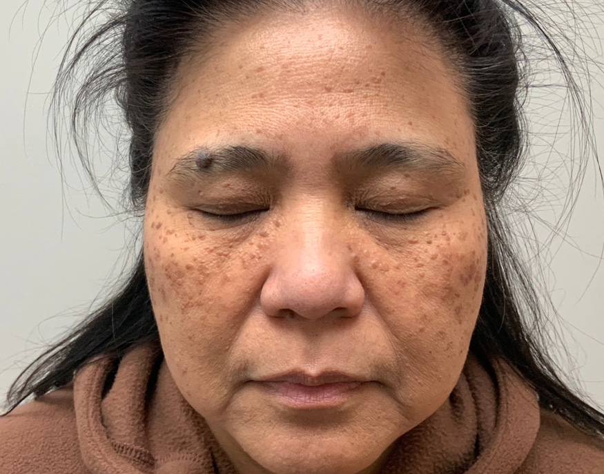 BEFORE
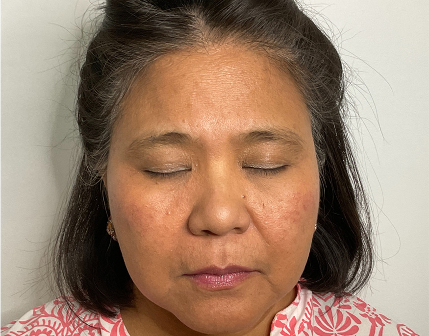 AFTER
AFTER
Skin lesions
Remove benign skin lesions with precision, minimizing scarring and promoting rapid healing.
Detailed Treatment Profiles
Detailed Treatment Profiles
Detailed information on each type of lesion and its removal approach.
Cherry Angiomas (Red Spots)
① How It Works
Cherry angiomas are small red growths formed by clusters of dilated blood vessels — generally benign and painless, but cosmetically bothersome. Laser treatment uses a wavelength specifically absorbed by hemoglobin in the lesion's blood vessels.
This generates heat that coagulates the vessels and triggers the body's gradual resorption of the lesion. Surrounding tissue is preserved through selective targeting. Most cherry angiomas can be effectively treated in a single session.
Key information
Anesthesia
Topical numbing cream
Invasiveness
Low
Duration
A few minutes
Recovery
Minimal
Discomfort
Low
Session
Generally 1 to 2
② What to Expect
Treatment is brief — most small angiomas are treated in seconds to minutes. A light tingling or warmth may be felt.
Mild redness and occasionally a small temporary crust may occur. The lesion gradually fades over a few weeks. Prior medical evaluation is mandatory to confirm the diagnosis.
Ideal for:
- Cherry angiomas on the face, trunk, or back
- Benign red vascular lesions
- Cosmetically bothersome red spots
③ Results
Most cherry angiomas can be reduced or eliminated after 1 to 2 sessions. Results are progressive — the lesion fades over several weeks as the body reabsorbs treated vessels.
Cherry angiomas can be reduced or eliminated.
Keratosis
① How It Works
Keratoses are thickenings of the skin's outer layer. Seborrheic keratoses are waxy or rough, brownish or light beige lesions, often appearing "pasted" onto the skin — linked to aging and genetics. Actinic keratoses are dry, rough patches linked to prolonged sun exposure.
For both types, laser ablation precisely vaporizes keratinic tissue in a controlled manner, resulting in more even skin post-healing.
Key information
Anesthesia
Local or Topical
Invasiveness
Low
Duration
Variable
Recovery
A few days
Discomfort
Low
Session
Generally 1 main session
② What to Expect
A local or topical anesthetic is applied before treatment.
A small crust may form after treatment and naturally detaches within a few days. Sun avoidance is recommended during healing.
Ideal for:
- Seborrheic Keratoses
- Actinic Keratoses
- Rough or thick growths
③ Results
Results are generally noticed after one session and complete healing.
The lesion is generally eliminated or significantly reduced
Uniform skin improvement
Skin Tags (Acrochordons)
① How It Works
Skin tags are small skin-colored growths attached by a thin stalk. Although benign, they can be cosmetically bothersome or irritated by clothing and jewelry.
Laser removal uses concentrated energy to cut the stalk at its base with high precision, leaving minimal impact on surrounding skin.
Key information
Anesthesia
Topical numbing cream
Invasiveness
Low
Duration
A few minutes
Recovery
A few days
Discomfort
Low
Session
Generally 1 session
② What to Expect
Anesthetic cream can be applied based on area sensitivity. Treatment takes seconds to minutes per lesion. Mild redness subsides quickly, and the area heals in a few days.
Ideal for:
- Skin tags on the neck, eyelids, armpits
- Cosmetically bothersome skin tags
- Lesions irritated by clothing or jewelry
③ Results
Results develop progressively over several weeks
Most skin tags are permanently removed in one session.
Healing is generally fast
Favorable aesthetic result
Moles (benign nevi)
① How It Works
Moles are clusters of pigmented cells that can be flat or raised and may evolve slowly over time. Before removal, a thorough medical evaluation is essential to confirm benign status and rule out suspicious signs requiring specialized dermatological care.
Once confirmed benign, certain moles can be removed by laser or another adapted technique.
Key information
Anesthesia
Local
Invasiveness
Low to moderate
Duration
Variable
Recovery
Variable
Discomfort
Low to moderate
Session
Generally 1 session
② What to Expect
Prior medical evaluation is mandatory. A local anesthetic is applied. Healing time varies based on depth and technique used.
Ideal for:
- Medically confirmed benign nevi
- Cosmetically bothersome moles
- Lesions in friction areas
③ Results
The mole can be removed with good aesthetic results
results depending on its characteristics and the chosen technique.
Ready to remove
your skin lesions?
Book your free medical consultation. Our physicians will evaluate your skin and recommend the approach best suited to your needs.